
09/07/2024
Share:
NEW YORK, July 9: A new 3D imaging model of early-stage embryos may enhance the success rates of in vitro fertilization (IVF), according to a team of scientists in China. This breakthrough could make it easier for women to conceive through IVF, a process responsible for about 2.5% of US births, equating to nearly 92,000 births in 2022.
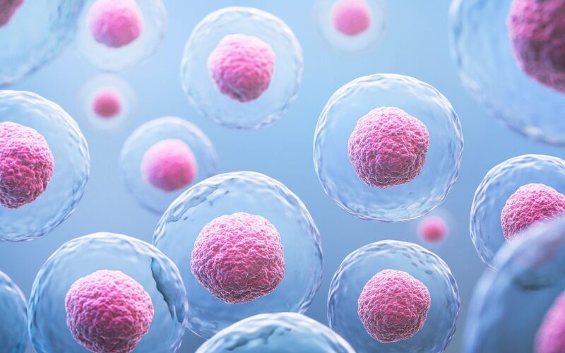
IVF often involves the creation of multiple embryos, making it challenging to identify which ones have the highest likelihood of resulting in a successful pregnancy. The Chinese researchers have developed a 3D model of blastocysts, embryos that are approximately five to six days old, which provides new insights into cell features linked to successful pregnancies.
“This study shows that the 3D shape of the blastocyst’s inner cell mass, its position, and how the surrounding cells are arranged can be important indicators of success, which we didn’t know before,” said Dr. Bo Huang, lead author and embryologist at the Reproductive Medicine Center of Tongji Hospital in China.
IVF involves collecting eggs from a woman and combining them with sperm in a laboratory to create embryos, which are then transferred into the uterus. Patients often have their embryos tested for genetic abnormalities before transfer. The success rate for genetically healthy embryos is between 60% and 65%, but these odds decrease with the woman’s age or if she has uterine conditions that impede embryo implantation.
The study included women under 40 with a thick uterine lining and no more than one previous failed embryo transfer. Using EmbryoScope+ technology, which monitors embryo development, researchers captured detailed images of 2,141 blastocysts. They compared their 3D model with fluorescence imaging and found it to be 90% accurate.
“Traditionally, the quality of blastocysts is assessed using 2D methods that lack depth and comprehensive indicators,” Huang explained. “Although some 3D methods exist, they aren’t practical or safe for clinical use. This study bridges that gap by introducing a clinically applicable 3D evaluation method and reveals previously unrecognized features of blastocysts.”
Dr. Huang’s research, published in the journal Human Reproduction, was presented at the annual meeting of the European Society of Human Reproduction and Embryology (ESHRE) in Amsterdam. He expressed hope that this technology will become a standard practice in IVF, providing new hope to patients struggling with infertility.